

Kidney Stones
Kidney stones can cause severe pain, urinary obstruction or infection, but not every stone requires surgery. Assessment helps establish where the stone is, whether it is causing a problem and which management pathway may be appropriate.
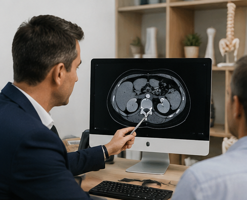
Infection behind urinary obstruction can require emergency drainage. Do not wait for a routine outpatient appointment if you are acutely unwell, have fever or chills with suspected obstruction, or are unable to pass urine.
What is a kidney stone?
A kidney stone is a solid deposit that develops from substances within the urine. Stones may remain inside the kidney or move into the ureter, the tube carrying urine from the kidney toward the bladder.
Kidney stones vary greatly in size, number, location and composition. Some are found incidentally and never cause symptoms. Others move, obstruct urine drainage or become associated with infection.
Those differences are important because a small uncomplicated stone, a stone lodged within the ureter and a large branching kidney stone can require very different management.
Explore the complete Kidney Stones & Endourology centre →
What can a kidney stone feel like?
Symptoms depend particularly on where the stone is located and whether it is moving or obstructing urine drainage.
Severe flank or back pain
Renal colic can produce sudden severe pain in the side or back, sometimes occurring in waves.
Pain travelling toward the groin
Pain can move toward the lower abdomen or groin as a stone progresses through the ureter.
Blood in the urine
A stone can produce visible blood in the urine or microscopic blood detected during testing.
Nausea or vomiting
Severe stone pain is commonly accompanied by nausea and may cause vomiting.
Urinary symptoms
Stones closer to the bladder can sometimes cause urinary urgency, frequency or discomfort.
No symptoms at all
Some kidney stones are found incidentally during ultrasound, CT or other imaging performed for another reason.
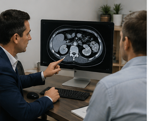
A CT shows much more than whether a stone is present
Imaging can determine where a stone is located, its approximate size and whether it is interfering with drainage from the kidney.
A non-contrast CT can also provide information about the total stone burden, urinary anatomy and stone density. These factors may influence whether surveillance, shock-wave treatment, ureteroscopy or PCNL is discussed.
Is the stone still in the kidney or has it entered the ureter?
This is one of the most important distinctions when understanding what may happen next.
Stone inside the kidney
Some renal stones remain stable and asymptomatic and can be monitored. Others grow, cause recurrent symptoms, become associated with infection or reach a size or location where active treatment may be more appropriate.
Learn about surveillance →Stone travelling toward the bladder
A stone that has entered the ureter can cause renal colic and obstruction. Some pass naturally, while others are unlikely to pass or require treatment because of persistent symptoms or obstruction.
Explore ureteric stones →Does your stone actually need treatment?
Stone size is important, but treatment decisions also depend on location, symptoms, obstruction, infection, kidney function, CT characteristics, previous treatment and individual circumstances.
Observation, shock waves, ureteroscopy or PCNL?
There is no single procedure that is best for every kidney stone. Each pathway addresses a different stone problem.
Observation
Selected stones may be monitored when immediate intervention is not required.
- Selected asymptomatic stones
- Follow-up imaging
- Monitor growth or obstruction
Shock-Wave Lithotripsy
Externally generated shock waves can fragment appropriately selected urinary stones.
- Suitability depends on stone factors
- CT density can be relevant
- Fragments pass afterwards
Ureteroscopy & Laser
Fine instruments can reach selected stones through the natural urinary tract for direct endoscopic treatment.
- Common ureteric stone pathway
- Flexible access to many renal stones
- Laser fragmentation where appropriate
PCNL
Direct keyhole access through the back is particularly important for selected larger and more complex renal stone burdens.
- Larger renal stone burdens
- Many staghorn stones
- Selected complex stones
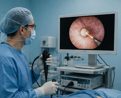
Modern stone treatment is usually minimally invasive
Many urinary stones can now be managed using endoscopic instruments passed through the urinary tract or with small-access techniques such as PCNL rather than conventional open stone surgery.
The best access route depends on where the stone sits, how much stone is present and the anatomy that must be navigated to clear it.
Some kidney stones need a more specific pathway
If your scan or medical history identifies a particular situation, these pages go beyond the general kidney-stone overview.
Blocked or Infected Stone
Why urinary infection behind an obstruction can require urgent drainage.
Explore urgent obstruction → ObservationAsymptomatic Kidney Stones
Understand when an incidentally detected renal stone may be monitored.
Explore surveillance → Complex stonesLarge & Complex Stones
Treatment planning for larger, multiple or difficult renal stone burdens.
Explore complex stones → Complex renal stoneStaghorn Kidney Stones
Assessment and planning for larger branching stones involving the kidney.
Explore staghorn stones → Stone compositionUric Acid Stones
Why urinary pH and stone composition can change management.
Explore uric acid stones → Special circumstanceKidney Stones in Pregnancy
Pregnancy changes imaging, observation and treatment considerations.
Explore pregnancy pathway → CT assessmentStone Density & Hounsfield Units
Understand what CT density can add to kidney stone treatment planning.
Understand HU → After treatmentResidual Kidney Stones
What remaining fragments can mean after previous stone treatment.
Explore residual stones → PreventionRecurrent Kidney Stones
Explore why stones recur and when metabolic assessment may help.
Explore prevention →Stone care does not always finish when the stone is removed
Recovery, residual fragments and recurrence prevention can become the next part of the pathway.
Ureteric Stents
A temporary stent may be used before or after selected procedures to maintain urine drainage between the kidney and bladder.
What to expect with a stent →Residual Stone Fragments
Follow-up imaging may show remaining fragments that require observation, further treatment or interpretation in the context of the original stone.
Learn about residual stones →Preventing Another Stone
Recurrent stone formers may benefit from stone analysis, blood tests and selected metabolic investigations such as 24-hour urine testing.
Explore 24-hour urine testing →What should I bring to a kidney stone appointment?
Existing investigations are particularly useful because treatment planning is often driven by the actual imaging rather than the radiology report alone.
Looking for the complete kidney stone service?
The Kidney Stones & Endourology centre brings together ureteric stones, treatment procedures, complex stones, stents, prevention and specialist stone pathways.
Kidney stone FAQs
Does every kidney stone need surgery?
No. Selected kidney stones can be safely monitored, and some stones that have entered the ureter can pass naturally. Treatment becomes more relevant when there is persistent pain, obstruction, infection, stone growth, threatened kidney function, substantial stone burden or a low likelihood of spontaneous passage.
What size kidney stone needs surgery?
There is no single measurement that automatically means a stone needs surgery. Size is considered together with location, symptoms, obstruction, infection, kidney function, anatomy, overall stone burden and individual circumstances.
Can a kidney stone pass by itself?
Stones pass through the ureter rather than directly from the kidney. Many smaller ureteric stones can pass spontaneously, although the likelihood varies particularly according to stone size and location. Persistent obstruction, infection or uncontrolled symptoms can change the management plan.
What is renal colic?
Renal colic describes the severe pain commonly produced when a stone moves into or obstructs the ureter. Pain is often felt in the flank or back and can travel toward the abdomen or groin.
Do I always need a CT scan?
Imaging is selected according to the clinical situation. CT provides detailed information about urinary stone size, location, obstruction and other characteristics, while ultrasound and other imaging may be appropriate in selected circumstances.
What is ureteroscopy?
Ureteroscopy uses a fine endoscope passed through the natural urinary tract to reach a stone within the ureter. Flexible instruments can also reach many stones within the kidney, where laser energy may be used to fragment the stone.
What is PCNL?
Percutaneous nephrolithotomy, or PCNL, creates a small access tract directly into the kidney through the back or side. It is an important treatment pathway for selected larger and more complex kidney stone burdens.
Will I need a ureteric stent?
Not every patient requires a stent. A temporary ureteric stent may be used when maintaining internal drainage between the kidney and bladder is considered useful before or after stone treatment.
When is a kidney stone an emergency?
Urinary obstruction combined with infection can require urgent treatment. Fever, chills, significant illness or deterioration with suspected obstruction, inability to pass urine, uncontrolled symptoms or concern about threatened kidney function should prompt urgent medical assessment rather than waiting for a routine appointment.
Why do kidney stones keep coming back?
Recurrence can be influenced by urine volume, urinary chemistry, stone composition, diet, medical conditions, medications, genetics, infection and urinary anatomy. Selected patients with recurrent stones may benefit from metabolic assessment.
Not sure which kidney stone pathway applies to you?
You do not need to decide which procedure you require before being assessed. Existing imaging, symptoms and investigations can be reviewed to determine whether observation or active treatment may be appropriate and which options fit the stone.
This information is general educational information and does not replace individual medical advice. Kidney stone investigation and treatment depend on factors including stone size and location, symptoms, urinary obstruction, infection, kidney function, anatomy, previous treatment and individual clinical circumstances. Seek urgent medical care for fever or significant illness associated with suspected urinary obstruction.