

Kidney Stones During Pregnancy
Kidney stones can occur during pregnancy and may cause severe flank pain, nausea, urinary symptoms or urinary obstruction. Investigation and treatment are different from routine stone care because both maternal health and the pregnancy need to be considered.

Ultrasound is generally the first imaging test and treatment is coordinated with obstetric care where intervention is required.
Infection behind an obstructed kidney can become serious. During pregnancy, persistent vomiting, difficulty passing urine, fever, chills or significant deterioration should not wait for a routine outpatient appointment.
Kidney stone assessment requires a different approach during pregnancy
The objective is still to identify obstruction, infection and stones that require treatment, but imaging, medications, anaesthesia and the timing of procedures all require additional consideration.
Imaging is different
Ultrasound is preferred initially because it does not use ionising radiation. MRI or selected low-dose CT may be considered when the diagnosis remains uncertain.
Pregnancy itself can dilate the kidney
Physiological hydronephrosis is common during pregnancy, which can make it more difficult to distinguish normal pregnancy-related dilation from obstruction caused by a stone.
Medication needs review
Pain relief, anti-nausea medication, antibiotics and any proposed stone-passage medication need to be selected according to gestation and the individual pregnancy.
Treatment is multidisciplinary
Where intervention is required, treatment planning may involve urology, obstetrics, anaesthesia, radiology and other pregnancy-care services.

How is a kidney stone diagnosed during pregnancy?
Symptoms alone cannot reliably establish whether a kidney stone is present. Assessment commonly includes urine testing, blood tests and imaging to look for a stone, urinary obstruction and infection.
Ultrasound — first-line
Kidney and urinary tract ultrasound is generally the preferred first imaging test. It can identify hydronephrosis and some urinary stones without exposing the pregnancy to ionising radiation.
MRI — second-line
Non-contrast MRI can help define the level and cause of urinary obstruction when ultrasound has not provided a sufficiently clear answer.
Low-dose CT — selected cases
CT provides greater diagnostic accuracy for urinary stones but uses ionising radiation. It is therefore generally reserved for selected situations where ultrasound and MRI have not provided the information needed to safely guide management.

Can a kidney stone pass naturally during pregnancy?
Yes. Many uncomplicated symptomatic stones can initially be managed without surgery when symptoms are controlled and there is no evidence that urgent intervention is required.
Observation is not simply “doing nothing”. The purpose is to allow a reasonable opportunity for spontaneous stone passage while continuing to assess maternal wellbeing, urinary drainage and signs of complications.
When might a kidney stone need treatment during pregnancy?
Intervention is considered when continuing observation is less safe or symptoms cannot be adequately managed. The treatment chosen depends on whether the immediate priority is drainage or definitive stone removal.
Observation & trial of passage
Appropriate for many uncomplicated stones when symptoms are controlled and there are no clinical indications for urgent intervention.
- Clinical follow-up
- Pregnancy-appropriate symptom management
- Monitoring for infection or persistent obstruction
Ureteroscopy
A fine telescope is passed through the urinary tract to directly reach a ureteric stone. The stone may be removed or fragmented when appropriate.
- No external skin incision
- Can avoid prolonged temporary drainage
- Performed with pregnancy-specific anaesthetic and procedural planning
Ureteric stent
A temporary internal stent can restore urine drainage from the kidney when obstruction needs to be relieved without immediately treating the stone.
- Internal drainage
- Can rapidly relieve obstruction
- May require exchange during pregnancy
Nephrostomy
A nephrostomy tube drains the kidney directly through the back and may be used when urgent decompression is required or a ureteric stent is unsuitable.
- Direct kidney drainage
- Useful in selected acute situations
- Requires external tube care
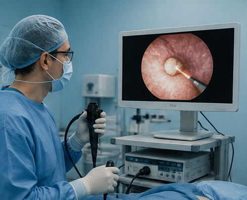
Ureteroscopy during pregnancy
Ureteroscopy has become an established treatment option for selected pregnant patients when a ureteric stone has not passed, symptoms remain difficult to control, obstruction is clinically significant or ongoing temporary drainage would otherwise be required.
The procedure is performed through the natural urinary tract. A ureteroscope is passed through the bladder into the ureter so the stone can be directly visualised and treated.
Ureteric stent or nephrostomy?
Both can decompress an obstructed kidney. Which approach is preferable depends on the clinical situation, urinary anatomy, infection, gestational considerations and local expertise.
Ureteric Stent
A thin internal tube passes from the kidney to the bladder to maintain urinary drainage.
- No external drainage bag
- Placed endoscopically
- Can cause urinary urgency, frequency or flank discomfort
- Pregnancy increases the tendency for stent encrustation
- Exchange may therefore be required if prolonged drainage is needed
Nephrostomy
A tube is placed directly through the back into the kidney to drain urine externally.
- Provides direct kidney drainage
- Can be used if retrograde stent placement is unsuitable
- Requires an external tube and drainage system
- Tube care and replacement may be required
- Choice is individualised according to the acute clinical situation
Which usual kidney stone treatments are different during pregnancy?
Shock-wave lithotripsy
Shock-wave lithotripsy should not be performed during pregnancy. Pregnancy is considered a contraindication to this treatment.
PCNL
Percutaneous kidney stone surgery is not a routine treatment pathway during pregnancy. Where considered at all, it requires individualised decision-making and appropriate expertise.
Ionising radiation
Ultrasound and MRI are preferred where they can answer the clinical question. CT or fluoroscopic imaging can still be used when genuinely required for safe diagnosis or treatment, with appropriate pregnancy-specific planning.
Does the trimester affect stone treatment?
Gestational age is one of several factors considered when planning imaging, anaesthesia and non-urgent intervention. The presence of infection, severe obstruction or another urgent clinical problem remains more important than simply waiting for a preferred trimester.
Conservative care where safe
Avoiding unnecessary intervention is generally desirable, but clinically important obstruction, infection or uncontrolled symptoms still require appropriate treatment.
Often preferred for non-urgent ureteroscopy
Where definitive ureteroscopy is required and timing can safely be planned, the second trimester is commonly favoured for non-urgent treatment.
Individualised planning
Treatment remains possible when clinically necessary, but obstetric factors, fetal monitoring requirements and proximity to delivery may influence the preferred strategy.
What about pain relief or medications to help the stone pass?
Medication choices during pregnancy require more care than for a non-pregnant stone patient. Suitable pain relief, anti-nausea treatment and antibiotics depend on the stage of pregnancy, allergies, other medical conditions and the clinical diagnosis.
Medical expulsive therapy is not something that should be started independently during pregnancy. Evidence for medications such as alpha-blockers in pregnancy is more limited than in the general stone population, and any proposed use should be reviewed by the treating urology and obstetric teams.
Do not assume a usual stone medication is pregnancy-safe.
If you are pregnant and being treated for renal colic, tell every treating clinician about the pregnancy and your gestational age before taking new prescription or over-the-counter medicines.

Can future stones be prevented?
Recurrent kidney stones may warrant further investigation, particularly if there have been several previous stone episodes, unusual stone types or other features suggesting an increased recurrence risk.
General prevention frequently includes maintaining appropriate hydration and a balanced diet, but pregnancy is not the time to begin an unnecessarily restrictive “kidney stone diet”.
More detailed metabolic assessment, stone analysis or 24-hour urine testing can be planned at the most appropriate time according to the stone history, pregnancy and postpartum pathway.
From suspected renal colic to a treatment plan
Assess symptoms
Establish the severity of pain, gestational age, vomiting, urinary symptoms, fever and previous stone history.
Investigate safely
Urine and blood testing are combined with pregnancy-appropriate imaging, usually beginning with ultrasound.
Observe or intervene
Uncomplicated stones may be observed. Infection, persistent obstruction or uncontrolled symptoms can change the pathway.
Plan follow-up
Confirm stone passage or drainage where necessary and consider further stone assessment after the acute episode has resolved.
Explore the relevant stone pathways
Kidney stones and pregnancy FAQs
Are kidney stones dangerous during pregnancy?
Many kidney stones can be managed safely without surgery. The situation becomes more concerning when there is urinary infection, persistent obstruction, uncontrolled symptoms, deterioration in kidney function or another complication. These circumstances require closer assessment and may require urinary drainage or stone treatment.
Can a kidney stone pass naturally while I am pregnant?
Yes. A trial of spontaneous passage is appropriate for many pregnant patients when symptoms can be controlled and there is no infection, significant deterioration or other indication for intervention.
What scan is safest for kidney stones during pregnancy?
Ultrasound is generally the first imaging test. MRI can be used when further imaging is required. Low-dose CT may be considered in selected situations when the diagnosis remains uncertain and obtaining an accurate diagnosis is important for clinical management.
Can an ultrasound miss a kidney stone?
Yes. Ultrasound is very useful for identifying urinary dilation and some stones, but it does not identify every ureteric stone. Normal pregnancy-related hydronephrosis can also complicate interpretation. Further imaging may occasionally be required.
Is MRI safe during pregnancy?
MRI does not use ionising radiation and is an accepted imaging modality during pregnancy when clinically indicated. For suspected urinary obstruction, non-contrast MRI is generally used; gadolinium contrast is not routinely required for stone assessment.
Can I have a CT scan while pregnant?
CT is not generally the first imaging test for suspected stones during pregnancy because it uses ionising radiation. However, necessary diagnostic imaging should not be withheld when the information is clinically important. Low-dose CT can therefore be considered in selected cases after the advantages and limitations of alternative imaging have been assessed.
Can kidney stone surgery be performed during pregnancy?
Yes. Ureteroscopy can be performed during pregnancy when definitive stone treatment is required. The decision is made with pregnancy-specific anaesthetic and obstetric planning. When treatment is non-urgent, the second trimester is commonly preferred.
Is anaesthesia safe if I need ureteroscopy?
If a procedure is clinically required, the anaesthetic plan is selected with consideration of gestational age, maternal health, the procedure and obstetric factors. Anaesthesia should be coordinated with the appropriate surgical and pregnancy-care teams rather than necessary treatment being avoided solely because the patient is pregnant.
Will I need a ureteric stent?
Not everyone needs a stent. A stent may be used to urgently drain an obstructed kidney, as temporary treatment while awaiting definitive stone management, or after selected ureteroscopy procedures.
Why may stents need changing more often during pregnancy?
Ureteric stents have a greater tendency to develop mineral encrustation during pregnancy. When a stent needs to remain in place for a prolonged period, a planned exchange strategy may therefore be required.
Can I have shock-wave lithotripsy while pregnant?
No. Pregnancy is considered a contraindication to shock-wave lithotripsy. Alternative management includes observation, urinary drainage or ureteroscopy depending on the clinical situation.
Can I take tamsulosin to help the stone pass during pregnancy?
Medical expulsive therapy during pregnancy requires individual medical assessment. The evidence base is more limited than for non-pregnant patients, and medications such as alpha-blockers should not be started independently without discussion with the treating urology and obstetric teams.
What should I do if I have fever and kidney stone pain while pregnant?
Seek urgent medical assessment. Fever, chills or significant illness associated with possible urinary obstruction can indicate infection behind a blocked kidney and may require urgent antibiotics and urinary drainage.
Guideline basis
- European Association of Urology. EAU Guidelines on Urolithiasis. Current online guideline. View guideline
- Pearle MS, Matlaga BR, Antonelli JA, et al. Surgical Management of Kidney and Ureteral Stones: AUA Guideline (2026), Part III. Journal of Urology. 2026;215(2):132–141. doi:10.1097/JU.0000000000004844.
- American College of Obstetricians and Gynecologists. Guidelines for Diagnostic Imaging During Pregnancy and Lactation. Committee Opinion No. 723. Reaffirmed 2026. View guidance
Have you developed a kidney stone during pregnancy?
Existing ultrasound, MRI or other investigations can be reviewed together with your symptoms, gestational age and obstetric history to determine whether continued observation, further imaging, urinary drainage or definitive stone treatment may be appropriate.