

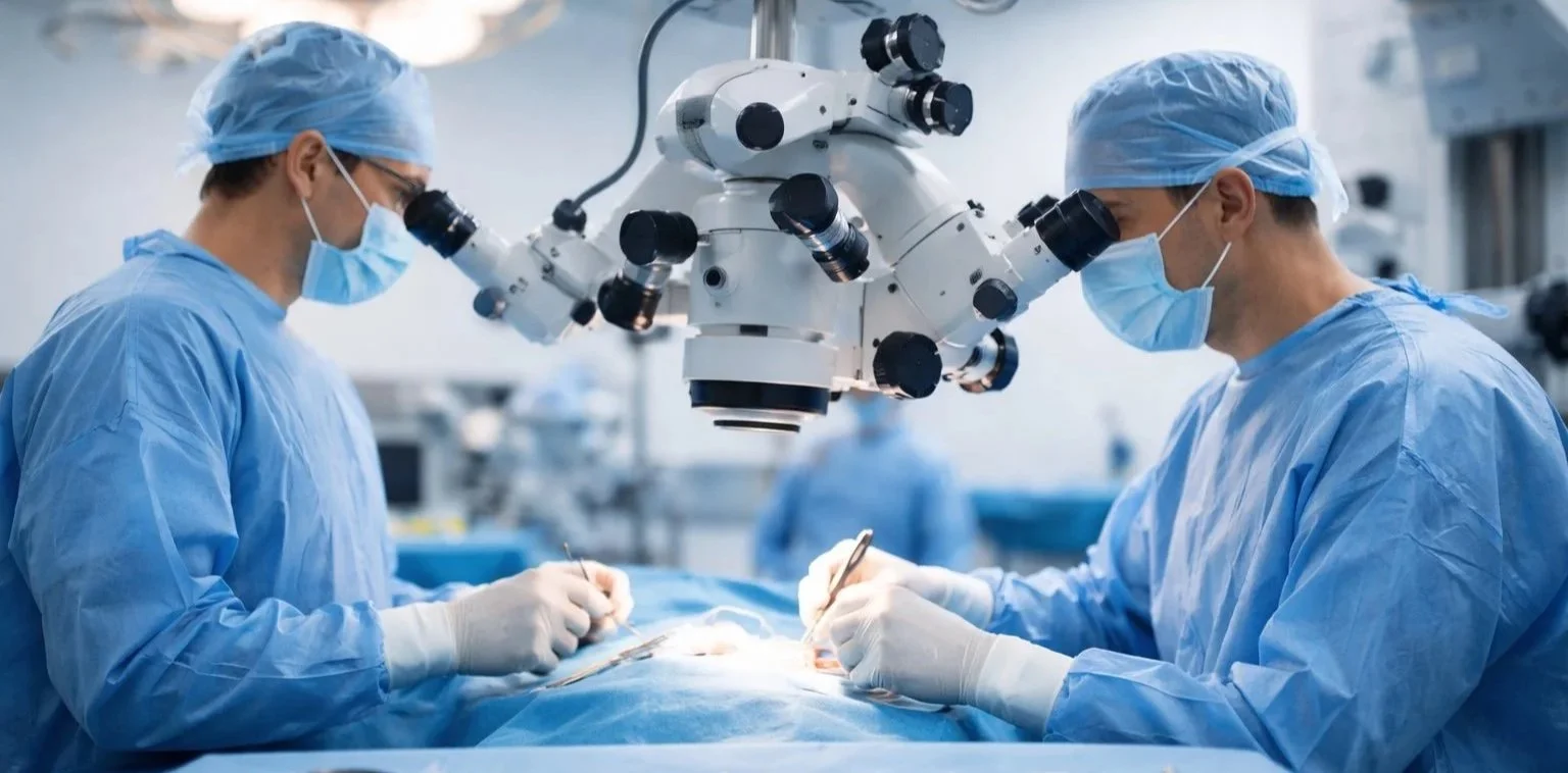
Scrotal and Testicular Surgery
Radical Orchidectomy (Removal of a Testicle)
What is a radical orchidectomy?
A radical orchidectomy is an operation to remove a testicle through a small incision in the groin (inguinal incision). The testicle and spermatic cord are removed as one unit.
This approach is different from removing a testicle through the scrotum. The groin approach is used because it is safest and most accurate when a testicular cancer is suspected.
Why is it performed?
Radical orchidectomy is most commonly performed when there is:
A suspicious testicular lump or mass on ultrasound
A strong concern for testicular cancer
Occasionally, a severely damaged or non-viable testicle (less common for “radical” approach)
It is both:
Diagnostic (confirms what the lump is under the microscope), and
Therapeutic (is the main first treatment for most testicular cancers)
What are the alternatives?
Depending on your situation, alternatives may include:
Observation / repeat ultrasound (only if imaging and clinical suspicion are low)
Testis-sparing surgery (uncommon; considered in very selected cases such as a small lesion, solitary testis, or bilateral tumours, and usually in specialist settings)
Further blood tests / imaging to guide planning
If cancer is suspected, biopsy through the scrotum is generally avoided because it can affect lymphatic drainage patterns and management.
Pre-operative preparation
Before surgery, you may have:
Scrotal ultrasound
Blood tests, including tumour markers (often AFP, β-hCG, LDH)
CT scan (sometimes before or after surgery, depending on timing)
Routine pre-anaesthetic assessment
Fertility preservation (important):
If you have not completed your family (or you’re unsure), ask about sperm banking before surgery—especially if:
You have reduced fertility already,
The other testicle is not normal,
You may need chemotherapy/radiotherapy later.
Medications:
Tell your team if you take blood thinners (warfarin, apixaban, rivaroxaban, clopidogrel, aspirin, etc.)
You’ll be advised what to stop and when.
Fasting:
You’ll receive instructions on when to stop food and liquids before your anaesthetic.
What happens on the day of surgery?
The surgery is usually done under general anaesthetic
A small cut is made in the groin
The testicle is delivered up from below and removed with the spermatic cord
The wound is closed with dissolvable stitches or stitches that need removal
A scrotal support may be recommended afterward
Length of surgery: often about 30–60 minutes (varies)
Hospital stay: commonly day surgery or overnight stay
Pain control
Most patients have manageable discomfort for several days.
Common pain relief options include:
Paracetamol and anti-inflammatory medication (if safe for you)
Short course of stronger pain relief if needed
Ice packs (wrapped, short periods) and supportive underwear can help
Recovery and activity
First 48 hours
Rest, keep the wound clean and dry
Wear supportive underwear
First 1–2 weeks
Expect bruising/swelling in the groin or scrotum
Gentle walking is encouraged
Avoid heavy lifting, strenuous exercise, and running
Driving
Usually once you can move comfortably and do an emergency stop safely (often several days; confirm with your surgeon and insurer)
Work
Desk/light duties: often 1–2 weeks
Heavy manual work: commonly 2–4+ weeks
Sex
Typically when comfortable and the wound is healing well (often 1–2 weeks), but follow your surgeon’s advice.
Wound care
Keep the dressing as directed
Showering is usually allowed after a set period; avoid soaking baths/pools until cleared
Watch for increasing redness, heat, swelling, or discharge
Risks and possible complications
All operations carry risk. Your surgeon will discuss your individual risk factors.
Common/expected
Bruising and swelling in the groin/scrotum
Temporary discomfort or numbness near the incision
Scrotal “emptiness” on that side
Less common but important
Bleeding/haematoma (blood collection)
Infection (wound infection or deeper infection)
Seroma (fluid collection)
Chronic groin discomfort or nerve irritation
Blood clots (DVT/PE) — uncommon but serious
Anaesthetic risks (varies by health and age)
Cancer-related considerations
Removing the testicle is usually the first step. Further treatment (surveillance, chemotherapy, radiotherapy, or surgery) depends on:
Pathology results
Tumour markers
Imaging findings
Fertility, hormones, and long-term effects
Fertility
Many men remain fertile with one healthy testicle.
Fertility may already be reduced in some men with testicular tumours.
If future fertility matters, discuss sperm banking early.
Testosterone
Most men maintain normal testosterone levels with the remaining testicle.
A minority may develop symptoms of low testosterone over time, such as:
Low energy, low mood, reduced libido, erectile changes
Reduced muscle mass, increased body fat
If symptoms occur, a simple blood test can check levels.
Testicular prosthesis (implant)
A testicular prosthesis can be placed to restore appearance and symmetry.
Options:
Inserted at the time of orchidectomy or as a later procedure
Not mandatory—some men prefer not to have one
Potential prosthesis risks:
Infection
Movement/position issues
Discomfort
Rarely, need for removal
Pathology results and follow-up
The removed testicle is sent for microscopy (histology).
Results typically return within several days to two weeks (timing varies).
At follow-up, your team may discuss:
Histology type (e.g., seminoma vs non-seminoma)
Tumour markers after surgery
Need for imaging and the best next step:
Surveillance (regular blood tests and scans)
Chemotherapy and/or radiotherapy
Additional surgery (selected cases)
Partial Orchidectomy (Testis-Sparing Surgery)
What is a partial orchidectomy?
A partial orchidectomy (also called testis-sparing surgery) is an operation where only the lump/tumour (and a small rim of surrounding tissue) is removed, while preserving as much of the testicle as possible.
It is different from a radical orchidectomy, where the entire testicle is removed through the groin.
Why is partial orchidectomy performed?
Partial orchidectomy is considered in selected situations, usually when preserving testicular tissue is important and safe. Common reasons include:
A small testicular mass where imaging and tumour markers suggest a lower risk of aggressive cancer
A benign tumour is suspected (e.g., Leydig cell tumour and other sex-cord stromal tumours)
A solitary testicle (only one testicle remains)
Bilateral tumours (masses in both testicles)
Strong fertility or testosterone preservation goals, especially when the other testicle is not functioning normally
Partial orchidectomy is typically done by surgeons and centres familiar with testis-sparing techniques and careful follow-up.
Who may not be suitable?
Partial orchidectomy may not be recommended when:
The mass is large or occupies a significant portion of the testicle
There are strongly suspicious features for typical testicular cancer
Tumour markers are significantly elevated or imaging suggests spread
The remaining testicular tissue is unlikely to be viable
There is concern that safe margins cannot be achieved
In these cases, radical orchidectomy may be safer and more definitive.
What are the alternatives?
Depending on your circumstances, alternatives may include:
Radical orchidectomy (standard first treatment if cancer is likely)
Active surveillance with repeat ultrasound (only when risk is low)
Biopsy/frozen section at surgery to guide whether partial or radical removal is needed (commonly part of the plan)
Sperm banking before any surgery or treatment (fertility preservation)
Your surgeon will explain the safest option for your diagnosis.
Pre-operative preparation
You may have:
Scrotal ultrasound (and sometimes MRI in selected cases)
Blood tests, including tumour markers (often AFP, β-hCG, LDH)
Routine blood tests and an anaesthetic assessment
Fertility preservation
If future fertility is important, ask about sperm banking before surgery, particularly if:
You have a single testicle,
Both testicles have lumps, or
Your semen parameters are already reduced.
Medications
Let your team know if you take blood thinners or antiplatelet agents.
Fasting
You’ll be given instructions before your anaesthetic.
What happens during the operation?
Partial orchidectomy is usually performed under general anaesthetic.
Typical steps
An incision is usually made in the groin (similar approach to radical orchidectomy) to protect oncological principles.
The testicle is delivered up and the lump is located.
Blood flow may be temporarily controlled (sometimes using a soft clamp) to reduce bleeding.
The lump is removed with a margin of tissue.
Often, the tissue is sent for urgent pathology (“frozen section”) during the operation.
Important:
If frozen section suggests a typical malignant germ cell tumour, your surgeon may recommend converting to a radical orchidectomy during the same operation for safety. This possibility should be discussed with you beforehand.
Operating time: often 60–120 minutes (varies)
Hospital stay: commonly day surgery or overnight
After the operation: pain control and wound care
Discomfort is common for a few days to a week.
Pain relief usually includes paracetamol ± anti-inflammatory medication (if safe for you).
Supportive underwear and short periods of ice packs (wrapped) may help.
Wound care
Keep the wound clean and dry as instructed.
Showering is usually allowed after a set period.
Avoid baths, pools, and spas until the wound is well healed.
Recovery and activity
First 48 hours
Rest and gentle walking around the house is encouraged.
First 1–2 weeks
Avoid heavy lifting, running, gym work, and straining.
Expect bruising and swelling of the groin/scrotum.
Work
Desk/light duties: often 1–2 weeks
Heavy manual work: commonly 2–4+ weeks
Driving
When comfortable and able to perform an emergency stop safely (check with your surgeon and insurer).
Sex
Usually once comfortable and healing is progressing (often 1–2 weeks), follow your surgeon’s advice.
Benefits of partial orchidectomy
Potential benefits include:
Preserving testosterone production
Preserving fertility potential (depending on baseline fertility and how much tissue remains)
Maintaining the appearance and feel of the testicle
Risks and possible complications
All surgery carries some risk. Your individual risk depends on your health and the details of your case.
Common/expected
Bruising, swelling, and discomfort
Temporary numbness or tenderness near the incision
Less common but important
Bleeding/haematoma (blood collection)
Infection
Seroma (fluid collection)
Chronic pain (nerve irritation or scarring)
Reduced testicular function in the operated testicle
Testicular atrophy (shrinkage due to reduced blood supply)
Recurrence or residual tumour (if malignant tissue remains or returns)
Need for further surgery, including later radical orchidectomy
Cancer-specific considerations
If the lesion is malignant, testis-sparing surgery may require:
Very close follow-up
Sometimes additional treatment (depending on tumour type, margins, and presence of precancerous changes)
Fertility and hormones (testosterone)
Fertility
Fertility after partial orchidectomy depends on:
baseline sperm quality,
the amount of healthy tissue preserved,
and whether further treatments are needed.
Sperm banking may still be recommended.
Testosterone
Preserving testicular tissue can help maintain testosterone production.
If symptoms of low testosterone occur (fatigue, low libido, mood changes), a blood test can check levels.
Pathology results and follow-up
The removed tissue is sent for detailed pathology. Results usually return within several days to two weeks.
Follow-up may include:
Wound check
Review of pathology
Repeat tumour markers and/or imaging where appropriate
A surveillance plan (clinical exams and ultrasound intervals if recommended)
Hemiscrotectomy for paratesticular tumours
What are paratesticular tumours?
Paratesticular tumours arise from structures next to the testicle, such as:
The spermatic cord
The epididymis
The tunica / coverings around the testicle
Scrotal soft tissues (fat, muscle, connective tissue)
Many paratesticular masses are benign, but some are malignant (cancerous), including soft tissue sarcomas (e.g., liposarcoma, leiomyosarcoma) and other rarer tumour types.
Why is hemiscrotectomy and radical orchidectomy recommended?
For suspected or confirmed malignant paratesticular tumours, the safest approach is usually complete removal with a clear margin of normal tissue around the tumour.
This operation is recommended when:
The tumour involves (or may involve) the spermatic cord and/or scrotal tissues
The mass is suspicious for sarcoma or other malignant tumour
The tumour is close to the scrotal skin or may have spread into scrotal tissues
The aim is to reduce the chance of local recurrence (the tumour coming back in the same area)
Hemiscrotectomy means removing the affected half of the scrotum (skin and underlying tissues as needed).
Radical orchidectomy means removing the testicle and spermatic cord, usually through a groin incision with a “high” cord tie.
These are often done together (en bloc) to give the best chance of complete tumour removal.
What are the alternatives?
Alternatives depend on the tumour type, location, and stage. They may include:
Local excision only (sometimes suitable for small, clearly benign lesions)
Different surgery based on specialist pathology (rare tumour types can change planning)
Radiotherapy and/or chemotherapy (often used in addition to surgery in selected cases)
Observation (generally only for lesions confidently assessed as benign)
Your surgeon will discuss why the recommended approach is best for your situation.
Pre-operative assessment and preparation
You may require:
Ultrasound and/or MRI of the scrotum/groin
CT scan of chest/abdomen/pelvis to check for spread (especially with suspected sarcoma)
Blood tests and routine pre-anaesthetic assessment
Biopsy:
In some cases, a needle biopsy is arranged before definitive surgery. In others, the mass is removed as part of the operation. The best approach depends on the suspected tumour type and imaging findings.
Fertility and sperm banking:
If you may want children in the future, ask about sperm banking before surgery, especially if:
You have only one functioning testicle, or
The other testicle is abnormal or has reduced function, or
You may need chemotherapy/radiotherapy later.
Hormones (testosterone):
Most men with a normal remaining testicle maintain normal testosterone. If the remaining testicle is not normal, testosterone monitoring and treatment options can be discussed.
Medications:
Tell your team if you take blood thinners (e.g., warfarin, apixaban, rivaroxaban, clopidogrel, aspirin). You will be given a plan for stopping/restarting them safely.
What happens during the operation?
This surgery is performed under general anaesthetic.
Typical steps
Groin incision (inguinal approach): the spermatic cord is controlled and divided “high” in the groin to reduce risk of tumour spread.
Radical orchidectomy: the testicle and spermatic cord are removed.
Hemiscrotectomy: the involved half of the scrotum is removed. The amount removed depends on:
tumour size and location
whether the tumour is close to or involves scrotal skin
the margin needed for safe clearance
Reconstruction/closure: the remaining scrotal skin is typically used to close the area.
If a larger area is removed, reconstructive techniques may be required (local flap or skin graft).
A drain may be placed to reduce fluid build-up and assist healing.
Operating time: varies (often 1–3 hours depending on complexity)
Hospital stay: commonly 1–2 nights (sometimes longer if reconstruction is needed)
After surgery: what to expect
Pain and swelling
Bruising and swelling of the groin/scrotum are common.
Pain is usually controlled with simple medications (paracetamol ± anti-inflammatories if safe) and occasionally stronger medication for a short time.
Dressings and drain
You may go home with a drain. The team will tell you:
how to care for it
when it will be removed (often within several days, but timing varies)
Support
Supportive underwear/scrotal support helps reduce discomfort and swelling.
Recovery and activity
First 48 hours
Rest, gentle walking is encouraged.
Keep dressings clean and dry as instructed.
First 2 weeks
Avoid heavy lifting, running, gym work, and straining.
Expect ongoing bruising and swelling.
Work
Desk/light duties: often 2 weeks
Physical work: commonly 4–6+ weeks (depends on wound size and reconstruction)
Driving
When you can move comfortably and safely perform an emergency stop (check with your surgeon and insurer).
Sex
Usually when comfortable and wounds are healing well (often 2–4 weeks), depending on extent of surgery.
Risks and possible complications
All surgery carries risks. Your surgeon will discuss your individual risk profile.
Common/expected
Pain, bruising, swelling
Temporary numbness or altered sensation around the groin/scrotum
Scarring and cosmetic change
Wound-related
Infection
Bleeding/haematoma
Seroma (fluid collection)
Delayed wound healing (more likely if larger hemiscrotectomy or reconstruction)
Wound breakdown (rare, but important)
Other risks
Chronic groin discomfort or nerve irritation
Inguinal hernia (uncommon)
Blood clots (DVT/PE) — uncommon but serious
Anaesthetic risks
Cancer-related
Positive margins (tumour cells at the edge of the specimen) may require:
further surgery, and/or
radiotherapy
Local recurrence can occur even after complete surgery, so follow-up is important.
Fertility, testosterone, and prosthesis options
Fertility
With one normal testicle, many men remain fertile.
If fertility is a priority, discuss sperm banking before surgery and whether semen testing is appropriate afterward.
Testosterone
Most men keep normal testosterone if the remaining testicle is healthy.
If symptoms of low testosterone occur (low energy, low libido, mood changes), blood tests can check levels and treatment can be discussed.
Testicular prosthesis
A testicular implant may be an option, but with hemiscrotectomy/reconstruction it may be:
best placed later once healing is complete, or
not recommended in some cases (e.g., if radiotherapy is planned or infection risk is higher).
Your surgeon will advise.
Pathology results and further treatment
The removed tissue is examined in a laboratory to confirm:
tumour type
grade (how aggressive it looks)
size
margin status (whether it was completely removed)
Results usually return within 1–2 weeks (timing varies).
Further treatment may include:
Surveillance (regular examinations and imaging)
Radiotherapy (commonly used for some sarcomas to reduce local recurrence risk)
Chemotherapy (selected cases depending on tumour type and stage)
Additional imaging and multidisciplinary team review

Scrotal Surgery for Lymphedema
What is scrotal lymphedema?
Scrotal lymphedema is long-term swelling of the scrotum caused by poor lymphatic drainage. Over time, the skin and tissues can become thickened, heavy, and prone to infection, leading to major problems with comfort, mobility, hygiene, urination, and sexual function.
Why might surgery be recommended?
Surgery is considered when scrotal lymphedema is:
Severe, heavy, and disabling
Causing recurrent infections (cellulitis), skin breakdown, or constant weeping
Making hygiene difficult or causing persistent chafing/rash
Associated with urinary issues (spraying, difficulty voiding standing) or sexual dysfunction
Not adequately controlled with conservative treatments (support garments, lymphedema therapy, skin care)
The goal of surgery is to remove diseased tissue and reconstruct a functional scrotum, improving quality of life.
What does the surgery involve?
Scrotal surgery for lymphedema is typically excisional/debulking surgery with reconstruction.
Depending on your anatomy and severity, the operation may include:
1) Removal of diseased scrotal tissue
Thickened lymphedematous skin and underlying tissue are removed.
The testes and spermatic cords are carefully protected.
2) Scrotal reconstruction
A new scrotum is created using remaining healthy scrotal skin where possible.
If insufficient healthy skin remains, reconstruction may require:
local tissue flaps, and/or
skin grafting (less commonly for the scrotum than the penis, but sometimes needed)
3) Penile management
If lymphedema has caused a buried penis or the penile skin is affected:
The penis may be “unburied”
Diseased penile skin may be removed
A skin graft may be used to cover the penile shaft
Not everyone needs penile reconstruction—your surgeon will explain what applies to you.
Benefits of surgery
Potential benefits include:
Reduced scrotal size and weight
Improved mobility and comfort
Easier hygiene and skin care
Fewer infections and less weeping/chafing
Improved ability to pass urine (especially if a buried penis is corrected)
Improved sexual function and body confidence (varies)
Pre-operative preparation
You may have:
Scrotal ultrasound (to assess testicles and exclude other issues)
Sometimes CT/MRI if there is concern about underlying obstruction, tumour, or anatomy
Routine blood tests and anaesthetic assessment
Optimising before surgery improves outcomes:
Weight management (if relevant)
Smoking cessation
Treating fungal rash/skin infection before surgery
Managing diabetes and other medical conditions
Lymphedema therapy:
Some patients benefit from pre-op input from a lymphedema therapist to improve skin condition and plan post-op maintenance.
Fertility and hormones:
If you have only one functioning testicle or reduced testicular function, discuss fertility/testosterone considerations.
Medications:
Tell your team about blood thinners/antiplatelets—these may need to be paused safely.
What happens on the day of surgery?
The operation is performed under general anaesthetic
A urinary catheter is commonly placed during surgery
Surgical drains are often placed to reduce fluid build-up
Compression/support garments and specialised dressings may be used
Hospital stay:
Varies by surgical extent:
Smaller reconstructions: sometimes a short stay
Larger reconstructions/skin grafting: often several days
Your surgeon will give you an expected length of stay.
After surgery: what to expect
Pain and swelling
Bruising, swelling and discomfort are expected initially.
Pain is usually managed with paracetamol ± anti-inflammatories (if safe), and sometimes short-term stronger pain medication.
Catheter and drains
A catheter may remain for a short period.
Drains remain until fluid output reduces; you may go home with a drain.
Dressings and wound care
Dressings may be bulky or involve special graft “bolsters” if grafting was performed.
Keep wounds clean and dry as instructed.
Your team will give specific showering instructions.
Recovery and activity
Recovery depends on the size of the surgery and whether grafting was required.
Typical guide:
First 1–2 weeks: rest, gentle walking, focus on wound care and swelling control
Avoid heavy lifting and strenuous exercise until cleared (often 4–6 weeks or longer for extensive reconstructions)
Return to work:
Desk duties: often 2–3 weeks
Physical work: often 6+ weeks
Driving: when you can move comfortably and perform an emergency stop safely
Sex: usually when wounds are well healed and comfortable (often several weeks; your surgeon will advise)
Risks and possible complications
All surgery carries risk. Important risks include:
Wound-related
Bleeding/haematoma (blood collection)
Seroma (fluid collection)
Infection
Delayed wound healing or wound breakdown
Unfavourable scarring
Swelling-related
Persistent swelling or recurrent lymphedema
Need for further procedures
If skin grafting is used (especially penile grafts)
Partial or complete graft loss
Tightness/scarring affecting erections or sensation
Change in skin colour/texture
General risks
Blood clots (DVT/PE)
Anaesthetic complications
Chronic pain or altered sensation
Your surgeon will discuss your individual risks based on your health and surgery type.
Long-term care after surgery
Even after successful surgery, ongoing care helps prevent recurrence:
Skin hygiene and moisturising
Weight management if relevant
Treat fungal rash early
Ongoing support garments if recommended
Early treatment of cellulitis
Some patients continue follow-up with a lymphedema therapist.
What is an epididymal cyst?
An epididymal cyst is a benign (non-cancerous) fluid-filled lump that forms in the epididymis—the coiled tube behind the testicle that stores and transports sperm.
If the cyst contains sperm, it is often called a spermatocele. Both are generally managed in the same way.
What causes epididymal cysts?
In most cases, the exact cause is unknown. They can occur at any age and are common. They are not usually linked to cancer.
Sometimes they may occur after:
Minor inflammation or infection
Previous trauma
Past surgery (less commonly)
Symptoms
Many epididymal cysts cause no symptoms and are found incidentally.
If symptoms occur, they may include:
A smooth lump above or behind the testicle
A feeling of heaviness or dragging discomfort
Scrotal ache (often mild)
Cosmetic concerns
Occasionally, discomfort during exercise or sex
Important: A new scrotal lump should be assessed to rule out other causes.
How is it diagnosed?
Diagnosis is usually based on:
Your history and physical examination
Scrotal ultrasound (commonly used to confirm the diagnosis and check the testicle)
Is it cancer?
Epididymal cysts are almost always benign and do not turn into cancer.
However, because testicular cancer can also present as a lump, it’s important to confirm the diagnosis—especially if the lump is new, growing, or attached to the testicle itself.
Management options
Treatment depends on size, symptoms, and your preferences.
1) Observation (most common)
If the cyst is small and not bothersome:
No treatment is needed
Many remain stable for years
Some slowly enlarge over time
A simple plan might include:
Periodic self-checks in the shower
Review if it enlarges or becomes painful
2) Pain management and supportive care
If there is mild discomfort:
Supportive underwear
Paracetamol or anti-inflammatory medication (if safe for you)
Avoiding activities that trigger pain
If pain is significant, your doctor may check for other causes (infection, inflammation, hernia, varicocele, testicular causes).
3) Aspiration (drainage with a needle)
This is not commonly recommended as routine treatment because:
The cyst often refills (high recurrence)
There is a risk of infection or bleeding
It can make later surgery more difficult due to scarring
In selected cases, aspiration ± sclerotherapy may be considered when surgery is not suitable.
4) Surgery (epididymal cyst excision)
Surgery may be considered if:
The cyst is large, growing, or uncomfortable
Symptoms affect daily activities, sport, or work
There is persistent pain clearly linked to the cyst
There are significant cosmetic concerns
Surgery: what to expect
Procedure: Epididymal cyst excision (often day surgery)
Anaesthetic: Usually general anaesthetic (sometimes regional/local in selected cases)
What happens
A small incision is made in the scrotum
The cyst is carefully separated from the epididymis and removed
The testicle is preserved
The wound is closed with dissolvable stitches
A dressing and scrotal support are applied
Recovery
Swelling and bruising are common for 1–2 weeks (sometimes longer)
Supportive underwear is usually recommended
Most people return to light activities within several days, and full activity over a few weeks
Potential impact on fertility (important)
Because the epididymis carries sperm, surgery can (rarely) affect fertility—especially if:
The cyst is large or complex
There are cysts on both sides
There is scarring or prior surgery/inflammation
If future fertility is important to you, discuss this with your surgeon before surgery.
Risks and possible complications
Most people do well, but risks include:
Common/expected
Bruising and swelling
Temporary discomfort or tenderness
A small scar
Less common
Infection
Bleeding/haematoma (blood collection)
Fluid collection (seroma)
Chronic pain or sensitivity
Recurrence or new cyst formation
Damage to the epididymis/vas deferens affecting fertility (uncommon)
Anaesthetic risks and blood clots (rare)
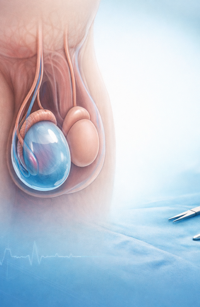
Hydrocele Repair (Hydrocelectomy)
What is a hydrocele?
A hydrocele is a collection of fluid around the testicle inside the scrotum. It commonly causes:
A painless scrotal swelling (often slowly enlarging)
Heaviness or discomfort, especially with exercise or at the end of the day
Cosmetic concerns
Hydroceles are usually benign (not cancer), but any new scrotal lump should be assessed to confirm the diagnosis.
Why is hydrocele repair performed?
Hydrocele repair may be recommended if the hydrocele:
Is large, uncomfortable, or getting bigger
Interferes with daily activities, exercise, sex, or clothing
Causes pain or significant heaviness
Raises uncertainty about the underlying testicle on examination/ultrasound
Recurred after aspiration (needle drainage)
Alternatives to surgery
Options depend on your symptoms, size of the hydrocele, and overall health:
Observation (watchful waiting)
Reasonable if it is small and not bothersome.
Aspiration (needle drainage) ± sclerotherapy
Fluid is drained with a needle; sometimes a medication is injected to reduce recurrence.
Often a higher chance of recurrence than surgery and may have infection/inflammation risks.
May be considered if you are not suitable for surgery.
Surgery (hydrocelectomy)
Usually the most definitive option with the lowest recurrence risk.
Pre-operative preparation
You may have:
Scrotal ultrasound (often done to confirm the diagnosis and check the testicle)
Routine blood tests and anaesthetic review (if needed)
Tell your team if you take:
Blood thinners (e.g., warfarin, apixaban, rivaroxaban, clopidogrel, aspirin)
Diabetes medications
Any supplements that increase bleeding risk (e.g., fish oil, high-dose vitamin E)
You will be given instructions about:
Fasting before surgery
Which medications to stop and when
Arranging someone to drive you home
What happens during hydrocele repair?
Hydrocele repair is usually performed under general anaesthetic (sometimes spinal or local in selected cases).
Typical steps
A small incision is made in the scrotum (occasionally in the groin depending on anatomy)
The hydrocele sac is opened, fluid drained, and the sac is removed or turned inside out (technique varies)
The testicle is checked
The wound is closed with dissolvable stitches
A dressing and supportive scrotal support are applied
Occasionally, a small drain is used (more common for large hydroceles)
Surgery duration: often 30–90 minutes
Hospital stay: usually day surgery
After surgery: what to expect
Pain and swelling
It is normal to have scrotal swelling and bruising.
Swelling can take weeks (sometimes longer for very large hydroceles) to settle.
Discomfort is usually manageable with paracetamol ± anti-inflammatory medication (if safe for you). Your surgeon may prescribe stronger pain relief briefly.
Dressings and support
You will usually be advised to wear supportive underwear/scrotal support for comfort and to reduce swelling.
Ice packs (wrapped, 10–15 minutes at a time) may help in the first 24–48 hours.
Wound care
Keep the dressing clean and dry as advised.
Showering is usually allowed after a set period; avoid baths, pools, and spas until healed.
Activity and recovery
Recovery varies with hydrocele size and your job/activity.
First 48 hours
Rest and gentle walking around the house is encouraged.
First 1–2 weeks
Avoid heavy lifting, running, gym work, and cycling.
Avoid straining; keep bowels regular (consider stool softener if needed).
Work
Desk/light duties: often 3–7 days
Manual/physical work: often 2–4 weeks
Driving
When you are comfortable, off strong pain medications, and can do an emergency stop safely.
Sex
Usually when comfortable and wounds are healed (often 2–3 weeks), follow your surgeon’s advice.
Risks and possible complications
All operations carry risk. Your surgeon will discuss your individual risk.
Common/expected
Bruising and swelling
Temporary discomfort or tenderness
Less common but important
Bleeding/haematoma (blood collection) — may rarely require drainage
Infection
Seroma (fluid collection)
Recurrence of the hydrocele
Chronic pain or sensitivity
Injury to structures near the testicle (rare), which could affect fertility in unusual cases
Anaesthetic risks and blood clots (uncommon but serious)