

Impacted Upper Ureteric Stones
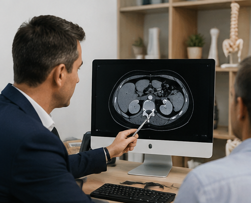
An impacted upper ureteric stone is a stone that has become lodged in the upper part of the ureter and has not progressed normally toward the bladder. Persistent impaction can cause obstruction and local ureteric inflammation, and may make treatment more technically challenging than for a freely mobile stone.
What does “impacted” actually mean?
Impacted does not simply mean “large”.
It describes a stone that has remained lodged at a particular point in the ureter rather than continuing to migrate toward the bladder.
When a ureteric stone remains in the same location, the surrounding ureter can become swollen and inflamed. The stone may become more closely embedded against the ureteric wall, while urine drainage from the kidney can remain partially or completely obstructed.
There is no single measurement on a CT scan that defines every impacted stone. The diagnosis is based on the overall picture, including the stone's position, duration, associated obstruction and findings encountered during treatment.
Why does the upper ureter matter?
The upper, or proximal, ureter is the section of ureter closest to the kidney. Stones in this area behave differently from stones that have travelled further down toward the bladder.
A small mobile stone may still progress naturally. However, an upper ureteric stone that remains fixed, causes persistent obstruction or repeatedly produces renal colic becomes less suitable for prolonged observation.
- Distance from the bladder
- Stone size
- Duration at the same location
- Degree of hydronephrosis
- Stone density
- Kidney function
- Previous treatment
- Associated infection
Why might an upper ureteric stone become impacted?
Impaction is usually the result of several factors acting together rather than one single cause.
Stone Size
Larger stones have greater difficulty travelling through the ureter and are generally less likely to pass spontaneously.
Ureteric Anatomy
Normal narrowing, angulation or an underlying area of ureteric narrowing can influence whether a stone progresses.
Time
A stone that remains in one position can produce increasing local swelling and inflammatory change around the stone.
Stone Shape
Irregular surface characteristics can influence how easily a stone moves within the ureter.
Previous Treatment
A persistent stone after observation or shock-wave treatment may require the treatment strategy to be reconsidered.
Ureteric Narrowing
Occasionally an underlying stricture or another anatomical factor contributes to a stone becoming lodged.
What does the CT scan need to show?
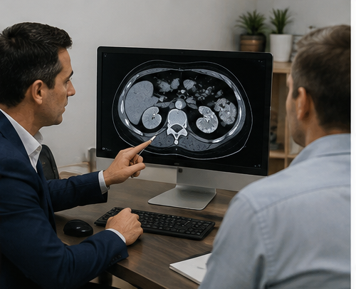
A CT scan does more than confirm that a stone is present. For an impacted upper ureteric stone, imaging can help determine both whether treatment is required and which approach is likely to be most appropriate.
Stone Size
The maximum dimensions and overall stone volume influence spontaneous passage and treatment.
Exact Position
The relationship between the stone, upper ureter and kidney helps plan endoscopic access.
Stone Density
Hounsfield units can provide information relevant to stone composition and shock-wave suitability.
Hydronephrosis
Swelling of the collecting system helps demonstrate the effect of obstruction on kidney drainage.
Kidney Anatomy
Collecting-system anatomy becomes particularly relevant when flexible or antegrade treatment is considered.
Other Stones
Additional renal stones may influence whether the treatment plan should address more than the ureteric stone.
Secondary Signs
Periureteric changes and the degree of obstruction can provide additional context when planning treatment.
Previous Imaging
Comparison with older scans can show whether the stone has remained fixed at the same site.
Does an impacted upper ureteric stone need treatment?
Not every upper ureteric stone requires immediate surgery. However, once a stone is persistently impacted, the likelihood of uncomplicated spontaneous passage may be lower and the consequences of ongoing obstruction become increasingly important.
Low Chance of Passage
Increasing stone size, persistent proximal location and failure to progress can shift the balance toward active stone removal.
Persistent Obstruction
Continued obstruction or hydronephrosis requires assessment even when pain is intermittent or has temporarily settled.
Persistent Pain
Recurrent renal colic or symptoms that cannot be adequately managed may be a reason to proceed to definitive treatment.
Kidney Function
Deteriorating renal function, bilateral obstruction or obstruction of a solitary functioning kidney requires particular attention.
Urinary Infection
Infection behind obstruction changes the priority from elective stone removal to urgent assessment and, where required, urinary drainage.
Patient Circumstances
Travel, occupation, access to emergency care, previous stone episodes and patient preference can all influence the timing of treatment.
How can an impacted upper ureteric stone be treated?
The best approach depends on the stone rather than simply whether the word “impacted” appears in a radiology report. Stone size, density, obstruction, anatomy, previous treatment and the ability to reach the stone all matter.
| Treatment | How it works | Where it may fit | Important considerations |
|---|---|---|---|
| Observation | Allows time for spontaneous passage. | Selected smaller stones without complications. | Less attractive when a stone remains fixed, obstruction persists, pain is recurrent or the chance of passage is low. |
| Shock-Wave Lithotripsy | External acoustic energy is focused on the stone to fragment it. | Selected proximal ureteric stones with favourable characteristics. | Success depends on stone size, density, visibility, body habitus, hydronephrosis and other anatomical factors. Additional treatment can be required. |
| Retrograde Ureteroscopy | A ureteroscope is passed through the bladder and upward to the stone. | Common definitive treatment for upper ureteric stones requiring removal. | Laser fragmentation, basket extraction and flexible ureteroscopy may be used depending on the stone and operative findings. |
| Staged Ureteroscopy | A stent is placed first and definitive ureteroscopy is performed later. | Useful when safe ureteroscopic access cannot initially be obtained. | Avoids forcing access through a tight or inflamed ureter and allows the ureter time to accommodate later instrumentation. |
| Antegrade / Percutaneous Treatment | The stone is approached from the kidney downward through a small percutaneous tract. | Selected large, impacted proximal stones or situations where retrograde access is unsuitable. | More invasive than routine retrograde ureteroscopy and reserved for selected clinical circumstances. |
| Urgent Drainage | A ureteric stent or nephrostomy restores urine drainage. | Infected obstruction or other situations requiring urgent decompression. | Drainage treats the immediate obstruction. Definitive stone removal is usually planned separately once appropriate. |
Ureteroscopy and laser treatment
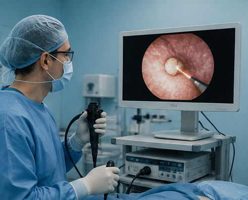
Ureteroscopy is commonly used when an upper ureteric stone requires definitive removal. A fine telescope is passed through the urethra and bladder and advanced upward through the ureter to the stone.
Laser energy can be delivered directly to the stone under vision. The aim may be to fragment the stone into removable pieces or reduce it into small fragments that can safely clear from the urinary tract.
For proximal stones, a flexible ureteroscope can be particularly useful. If the stone migrates upward into the kidney during treatment, flexible instruments may allow it to continue to be treated rather than abandoning the procedure.
What happens during treatment?
Reach the Stone
A ureteroscope is advanced carefully through the bladder and ureter toward the level of impaction.
Assess the Ureter
The degree of swelling, narrowing and stone impaction becomes clearer under direct endoscopic vision.
Treat the Stone
Laser lithotripsy can fragment the stone and selected pieces may be removed with an endoscopic basket.
Protect Drainage
A temporary ureteric stent may be placed when swelling, ureteric irritation, residual fragments or operative findings make additional drainage helpful.
Why might the operation be stopped and a stent inserted?
Occasionally the ureter is too narrow, inflamed or difficult to access safely during the first procedure.
In this situation, the safer option may be to insert a temporary ureteric stent rather than force instruments past a resistant area. The stent maintains drainage and can allow the ureter to become more accommodating before a later ureteroscopy.
This does not necessarily represent a failed operation. A staged approach can be a deliberate safety strategy when the anatomy encountered during surgery differs from what could be predicted from imaging alone.
Ureteric Stents – What to Expect →
What if the impacted upper ureteric stone is large?
Large impacted proximal ureteric stones can represent a different technical problem from a small mobile ureteric stone.
Retrograde ureteroscopy remains an important option, but treatment may become more difficult as stone size and impaction increase. Selected large proximal stones can also be approached from above through the kidney using an antegrade or percutaneous technique.
Current European guidance recognises percutaneous antegrade removal as an option in selected large impacted proximal ureteric stones, particularly when the collecting system is dilated or the stone cannot be managed effectively by the usual retrograde route.
- Large proximal stone burden
- Significant hydronephrosis
- Difficult retrograde access
- Previous unsuccessful treatment
- Associated renal stones
- Individual urinary anatomy
Can an impacted upper ureteric stone be treated with shock waves?
Shock-wave lithotripsy can treat selected upper ureteric stones without passing an endoscope into the urinary tract. External acoustic energy is focused onto the stone, with the goal of breaking it into fragments that can pass naturally.
The fact that a stone is in the upper ureter does not automatically make it suitable for shock-wave treatment. Success is influenced by several factors, including stone size, density, visibility, skin-to-stone distance and the degree of urinary obstruction.
When a stone is firmly impacted, has been present for a prolonged period or is associated with significant obstruction, ureteroscopic treatment may offer a more direct method of dealing with the stone.
Factors that can influence SWL suitability
- Stone size
- Stone density on CT
- Ability to accurately target the stone
- Skin-to-stone distance
- Degree of hydronephrosis
- Stone composition
- Urinary anatomy
- Previous SWL response
- Patient preference and clinical circumstances
What if the obstructed kidney is infected?
An infected obstructed urinary system is different from routine elective stone treatment. When significant infection or sepsis is present behind a blockage, the immediate goal is usually to restore urinary drainage and treat the infection.
Drain the Kidney
A ureteric stent or nephrostomy can restore drainage from the obstructed collecting system.
Treat the Infection
Antibiotic treatment and microbiological assessment are undertaken according to the clinical situation.
Treat the Stone Later
Definitive stone removal is generally planned once infection and systemic illness have appropriately resolved.

Will I need a ureteric stent?
Not every ureteroscopy requires a stent. However, stent use can be more relevant after difficult or impacted stone treatment because the ureter around the stone may be swollen or irritated.
A temporary stent may be used to:
- Maintain drainage while ureteric swelling settles
- Protect drainage after difficult instrumentation
- Allow stone fragments to pass
- Support healing following ureteric irritation or injury
- Prepare the ureter for a planned second procedure
The decision is usually based on the actual operative findings rather than the CT appearance alone.
Read the Ureteric Stent Guide →Why can impacted stones be more challenging to treat?
The ureter surrounding an impacted stone may have been exposed to prolonged pressure, obstruction and inflammation. This can affect both access to the stone and the condition of the ureteric wall.
Local Swelling
Oedema around the stone can make the true lumen of the ureter harder to identify or navigate safely.
Reduced Working Space
A tightly impacted stone can leave less room for instruments and stone fragments than a mobile ureteric stone.
Stone Migration
Proximal stones can move back into the kidney during fragmentation, sometimes requiring flexible ureteroscopy.
Ureteric Injury
Careful technique is particularly important where the ureteric wall is inflamed or closely adherent to the stone.
Residual Stone
Complete clearance may not always be appropriate during one operation if access, visibility or ureteric safety becomes limiting.
Later Narrowing
Significant inflammation or ureteric injury can occasionally contribute to later ureteric narrowing, which may influence follow-up in selected cases.
What follow-up might be needed?
Follow-up depends on the stone, degree of previous obstruction, treatment performed and operative findings. Imaging may be used to confirm stone clearance and that the kidney is draining appropriately.
Stone Clearance
Assess whether clinically significant fragments remain.
Kidney Drainage
Confirm that hydronephrosis and obstruction have resolved where appropriate.
Stent Removal
Remove any temporary ureteric stent according to the planned timeframe.
Stone Prevention
Recurrent stone formers may benefit from stone analysis and metabolic assessment.
Related information
Ureteric Stones
Symptoms, spontaneous passage, obstruction and the general treatment pathway for stones within the ureter.
Explore ureteric stones →Ureteroscopy & Laser Surgery
Learn how endoscopic instruments and laser energy are used to treat selected ureteric and kidney stones.
Explore ureteroscopy →Shock-Wave Lithotripsy
Understand when externally generated shock waves may be suitable for urinary stones.
Explore SWL →Stone Density & Hounsfield Units
Learn how CT stone density can influence treatment selection and the likely effectiveness of shock-wave treatment.
Understand stone density →Medical Expulsive Therapy
Understand when observation and medication may have a role while a ureteric stone is given time to pass.
Explore MET →Blocked or Infected Kidney Stone
Understand why infection combined with urinary obstruction can require urgent kidney drainage.
Learn about infected obstruction →Ureteric Stents – What to Expect
Why a temporary stent may be inserted, what symptoms it can cause and how it fits into stone treatment.
Read the stent guide →Kidney Stone Treatment Options
Compare observation, spontaneous passage, SWL, ureteroscopy, PCNL and urinary drainage.
Compare treatments →Kidney Stones & Endourology
Return to the complete kidney stone pathway including procedures, prevention and complex stone conditions.
Kidney stone hub →Impacted upper ureteric stone FAQs
What is an impacted upper ureteric stone?
It is a stone lodged in the proximal or upper ureter that has remained fixed rather than continuing to move toward the bladder. Prolonged impaction can be associated with obstruction, swelling and inflammation of the surrounding ureter.
Is an impacted stone the same as an obstructing stone?
Not exactly. A newly arrived stone can obstruct urine flow without being chronically impacted. Impaction generally refers to a stone that has remained lodged at one location, often with local ureteric inflammatory change.
Can an impacted ureteric stone still pass naturally?
Some stones may still pass, particularly if they are small and there are no complications. However, a stone that remains fixed, is larger, causes persistent obstruction or produces recurrent symptoms becomes less suitable for prolonged observation.
How long can an upper ureteric stone be left?
There is no single safe timeframe for every patient. Stone size and location, symptoms, degree of obstruction, infection and kidney function determine whether continued observation remains appropriate.
Is ureteroscopy usually used for an impacted stone?
Ureteroscopy is a common definitive treatment when an upper ureteric stone requires active removal. Laser energy can be used to fragment the stone and selected pieces can be removed endoscopically.
Can shock-wave lithotripsy treat an impacted upper ureteric stone?
Shock-wave treatment may be appropriate for selected proximal ureteric stones, but suitability depends on stone size, density, targeting, body habitus, obstruction and other factors. A firmly impacted stone may favour a more direct endoscopic approach.
What happens if the surgeon cannot get past the stone?
The safest strategy may be to avoid forcing access. A ureteric stent can sometimes be inserted and definitive ureteroscopy performed at a later procedure once the ureter has had time to accommodate instrumentation.
Will I need a stent after treatment?
Not everyone requires a stent. It may be particularly useful after difficult stone treatment when there is ureteric swelling, irritation, residual fragments, concern regarding drainage or a planned second procedure.
Can an impacted ureteric stone damage the kidney?
Persistent urinary obstruction can affect kidney function in some circumstances. The degree and duration of obstruction, whether both kidneys are affected and baseline kidney function are therefore considered when deciding how urgently a stone should be treated.
When is an impacted ureteric stone an emergency?
Urinary obstruction associated with infection can be a urological emergency. Fever, chills or rigors, significant illness, difficulty passing urine or deterioration with suspected obstruction should prompt urgent medical assessment.
Can a very large impacted upper ureteric stone be approached through the kidney?
Yes. In selected large impacted proximal stones, particularly when retrograde ureteroscopic access is difficult or unsuitable, an antegrade or percutaneous approach through the kidney can be considered.
If my pain has stopped, has the stone passed?
Not necessarily. Symptoms can settle despite a stone remaining within the ureter. Follow-up imaging may therefore be appropriate in selected patients to confirm stone passage and resolution of obstruction.
Have you been told your upper ureteric stone is impacted?
Existing CT imaging can be reviewed to assess the stone's size, position and density, whether the kidney remains obstructed and whether observation, ureteroscopy, shock-wave treatment or a more complex stone pathway may be appropriate.
Medical information: This information is general educational information and does not replace individual medical advice. Management of an impacted ureteric stone depends on stone size and location, duration, symptoms, obstruction, infection, kidney function, urinary anatomy, previous treatment and individual clinical circumstances. Seek urgent medical attention for fever, chills or significant illness associated with suspected urinary obstruction.
Clinical references
- European Association of Urology. EAU Guidelines on Urolithiasis. Current online guideline.
- Pearle MS, Matlaga BR, Antonelli JA, et al. Surgical Management of Kidney and Ureteral Stones: AUA Guideline (2026) Part I: Evaluation and Treatment of Patients With Kidney and/or Ureteral Stones. J Urol. 2026;215(2):113–123.
- Pearle MS, Matlaga BR, Antonelli JA, et al. Surgical Management of Kidney and Ureteral Stones: AUA Guideline (2026), Parts II and III. J Urol. 2026;215(2).